Thoracic outlet syndrome

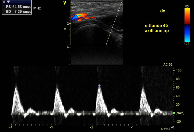
Ultrasound of the subclavian artery and also distal part of axillary artery in order to exclude pectoralis minor compression. Valuable technique to supplement MR or CT imaging, with the advantage of image acquisition while the patient is in a seated position and during arm elevation. Cessation of blood flow or significant increase in peak systolic velocity is indicative of arterial TOS.

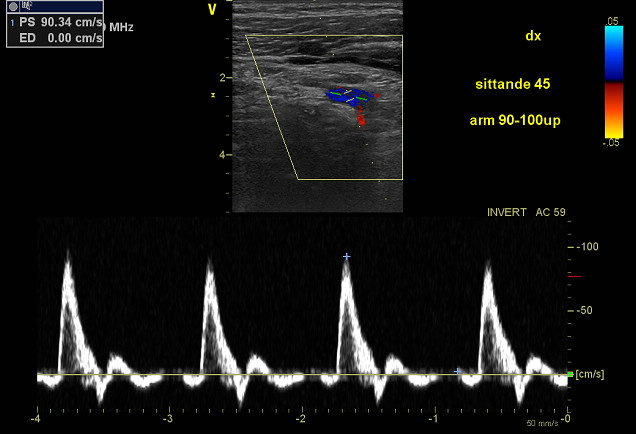
Subclavian artery viewed from infraclavicular position. No signifacant flow variation. Left image, arm down. Right image, arm up.

Distal part of axillary artery. No significant flow variation. Left image, arm down. Right image, arm up.

Thoracic outlet syndrome

Ultrasound of the subclavian artery and also distal part of axillary artery in order to exclude pectoralis minor compression. Valuable technique to supplement MR or CT imaging, with the advantage of image acquisition while the patient is in a seated position and during arm elevation. Cessation of blood flow or significant increase in peak systolic velocity is indicative of arterial TOS.
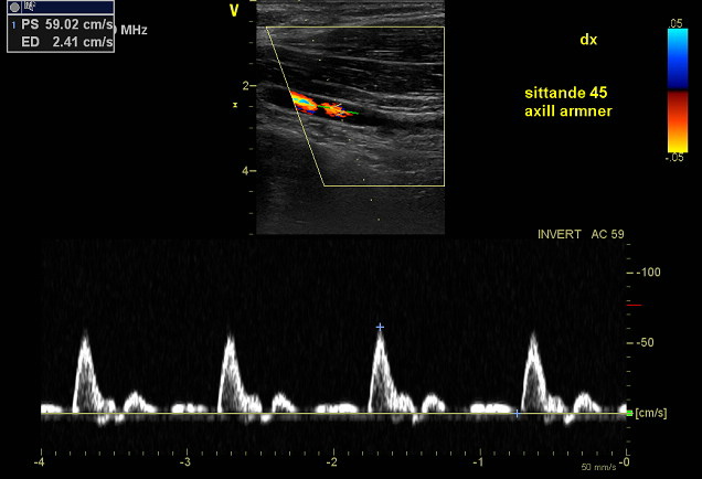
Subclavian artery viewed from infraclavicular position. No significant flow variation. Upper image, arm down. Lower image, arm up.

Distal part of axillary artery. No significant flow variation. Upper image, arm down. Lower image, arm up.

Stäng meny